Proceedings · Session S-365 · filed October 13, 2026
Lab Technology & MethodsSession paper
Human Prion Cell Model Cuts Assay Times from Years to Weeks
UCL's EKV cells propagate infectious human sCJD prions indefinitely, with assay sensitivity comparable to mouse bioassays — in weeks instead of years.
By Rebecca Stone4 min read871 words
Summary
- UCL researchers report EKV cells, the first scalable system propagating infectious human sCJD prions in dividing cells (PNAS).
- The Human Prion Assay built on EKV cells matches gold-standard mouse bioassay sensitivity and cuts timelines from years to weeks.
- ICSM18 antibody produced dose-dependent clearance of human PrPSc in infected EKV cells; an isotype control did nothing.
- SISSA researchers identified ARN1468, a serpin inhibitor that reduced prion load in cells; low bioavailability ruled out mouse studies.
- Only four vCJD cases have ever been reported in the United States, all linked to exposure abroad.
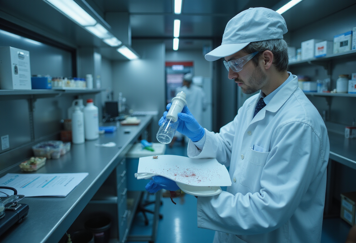
Researchers at University College London have grown infectious human prions in dividing cells for the first time in a scalable system, cutting the timeline for measuring infectivity, in their words, "from years to weeks." Parmjit Jat, PhD, professor of molecular biology at UCL, and colleagues published the work in PNAS, reporting EKV cells: a humanized mouse-cell line that supports robust, indefinite propagation of sporadic Creutzfeldt-Jakob disease (sCJD) prions — the most common human prion disease, which is always fatal.
For R&D managers in biomanufacturing and drug discovery, the advance addresses a bottleneck that has shaped budgets and portfolio decisions for decades: human prions resisted propagation in dividing cells, so infectivity studies leaned almost entirely on slow, costly animal bioassays.
Why propagation in cells matters
Prions are misfolded proteins — not viruses or bacteria — that convert healthy proteins into the same pathological shape, causing neuronal cell death. Jat and co-authors write that research into human prion biology and therapeutics "has been severely constrained by the inability to propagate human prions in dividing cells," leaving scientists dependent on "costly and slow animal bioassays."
The team reports that EKV cells reliably generate high-titer infectious sCJD prions. Lysates from those cells triggered lethal prion disease in humanized mice that was clinically and neuropathologically indistinguishable from disease caused by sCJD-infected human brain tissue.
From the cell line, the researchers built the Human Prion Assay (HPA), which quantifies sCJD infectivity with sensitivity they report as comparable to the gold-standard mouse bioassay. That reported equivalence, not superiority, is the measured claim; the decisive gain is turnaround time.
What does the platform change for screening?
The authors point to a concrete translational failure as motivation: earlier therapeutic screens built on murine prions identified compounds such as quinacrine that later failed in human trials. A cell-based system allows candidate testing against genuine infectious human prions from the start.
As a proof of platform, Jat's team dosed infected EKV cells with ICSM18, an anti-prion protein monoclonal antibody previously shown to work against nonhuman prions. The result: potent, dose-dependent clearance of human PrPSc (the pathological, misfolded form of prion protein) in infected cells. An isotype control antibody did nothing.
A renewable, cell-based model also makes experiments more reproducible and scalable. High-throughput screening of small-molecule inhibitors becomes experimentally realistic — more compounds, more concentrations, earlier rejection of failures.
A parallel strategy: targeting the machinery around prions
Separately, the laboratory of prion biology at the Scuola Internazionale Superiore di Studi Avanzati in Italy took a different route, published in the Journal of Medicinal Chemistry. Most strategies aim to block conversion of normal cellular prion protein, PrPC, into PrPSc. The Italian team instead targeted the biology surrounding accumulation.
Observing that the protease inhibitor SERPINA3 (SerpinA3n) is upregulated during prion disease, they identified a small molecule, ARN1468, that inhibits those serpins and reduced prion load in chronically infected cells. Low bioavailability ruled out in vivo studies in prion-infected mice. The researchers nonetheless call it "a novel and effective approach to the treatment of prion disease" — proof of concept that a drug can target cellular machinery rather than the prion protein itself. That claim rests on cell data alone.
What stays unchanged for manufacturers?
Biomanufacturing faces a parallel measurement problem, and the new cell assay does not replace it. Alison Armstrong, PhD, global head of scientific and regulatory consultancy at BioReliance Contract Testing Services, Merck KGaA, Darmstadt, explains: "Prions are infectious forms of naturally occurring proteins that can alter healthy proteins and lead to neuronal cell death." They present, she adds, "a potential health risk through animal-to-human transmission, although human-to-human transmission is rare."
"Because methods for directly detecting prions in raw materials are limited by sensitivity, prion-clearance studies have become an industry-standard approach for demonstrating that downstream purification steps can remove or inactivate transmissible spongiform encephalopathy agents," Armstrong says. A typical TSE risk assessment evaluates animal species, tissue source, country of origin, tissue classification, and sourcing from countries with negligible or controlled BSE risk.
Contract labs still anchor this work in older tools. ViruSure, staffed by veterans of the BSE crisis of the 1990s, uses the hamster-adapted 263K scrapie strain, quantitative Western blot assays, and in vivo bioassays to determine prion titer. Texcell runs spiking studies in scaled-down process vessels to track removal at each step.
The measured-versus-projected ledger
What the sources measure directly: EKV propagation of sCJD prions; disease induction in humanized mice indistinguishable from brain-tissue-derived infection; HPA sensitivity comparable to mouse bioassay; dose-dependent PrPSc clearance by ICSM18 in cells; reduced prion load in cells from ARN1468.
What remains projection: faster drug discovery timelines, high-throughput screening outcomes, and any therapeutic effect in patients. The distance between clearing PrPSc in a dish and treating a rapidly progressive, fatal neurodegenerative disease remains large, and the new cell assays will complement rather than replace animal and validation studies.
The practical significance for portfolio planning is nonetheless concrete: a system that propagates, quantifies, and experimentally clears genuine human sCJD prions shortens the experimental loop from years to weeks, letting laboratories challenge candidate treatments against the real pathogen at a scale animal bioassays never permitted.
via pnas.org (Original)
Filed under
- prion-disease
- cell-based-assay
- drug-discovery
- high-throughput-screening
- creutzfeldt-jakob-disease
More from Rebecca Stone
Show full bio
Market editor covering regulation, policy and their impact on research and development at Hypothesis Wire.
190 articles
References
- Baylor Team Couples Proteomics and AI to Degrade VAV1
- Talus Bio Releases Structure-Free AI Model for Disordered Proteome
- Spatial Biology Vendors Push From Discovery Bench to Clinical Assay
- Stem cell entrepreneurs lobby FDA for regulatory path, citing peptide precedent
- CRISPR-Transposon Platform Maps Essential Phage Genes Genome-Wide